Experience a versatile ultrasound solution for the modern healthcare environment.
The X-CUBE i9 offers optimum mobility, intuitive workflow, intelligent clinical applications, and efficient
diagnostic solutions.
Innovative healthcare professionals can achieve new heights in patient satisfaction
with best-in-class imaging clarity and accuracy to confidently support each critical diagnosis with
maximum viewing and data interpretations.

X-CUBE i9 frees you from time and space constraints
and helps you focus more on your patients. Long-lasting batteries and dual transducers provide flexibility in the examination
- Dual Transducers
- 2 active transducer connectors
(Second connector optional)
Double Up Your Efficiency
With dual batteries and transducers


- Dual Batteries
- 2 built-in batteries
(Second battery optional)

- Long-lasting dual batteries
- Standby mode: Max 100 hrs.
Continuous scan: Max 60 min.


- Easy Control Cart
- Easy adjustment of height: 848 ~ 998mm
Smooth and silent gas lift cylinder
Excellent Imaging Quality For Versatile Diagnostic Performance
Powered by X+ Architecture, innovative imaging algorithms
and efficient diagnosis workflow to effectively support the diverse range of clinical settings with crystal clear imaging quality.
The ultrasound acquisition process enhanced by the signal obtained with the X+ Crystal Signature, a highly sensitive broadband transducer and the high-resolution images generated by the X+ FIT technology, a large-capacity dataprocessing, provide a high-resolution image.


Expand Flexibility And Adaptability
X-CUBE i9 frees you from time and space constraints and helps you focus more on your patients. Long-lasting batteries and dual transducers provide flexibility in the examination and
the intelligent options help the fast examination and the guide function, which reduces the dependency on operators and maximizes efficiency.


X+ Assistant
Keystrokes have been reduced by more than 50% compared to conventional use, reducing examination time. Optimal scanning protocols are registered according to application-specific guidelines and users can optimize.

X+ Compare
Users are able to import previously acquired images from a PACS or hard disk and compare with the current image in real-time scan and in E-view mode for reviewing.
* X+ Compare supports ultrasound studies only.

Power Preset
Users can load a system preset saved in advance with a single button click. By using these quick and easy presets, users can shorten the imaging set up time.
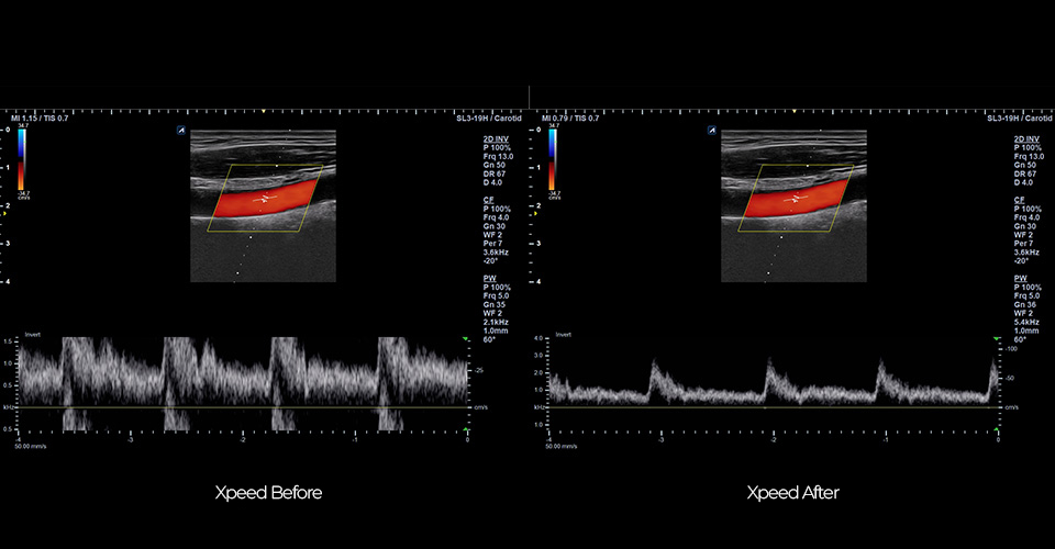
XpeedTM
Simply press the Xpeed™ button once to quickly optimize images in 2D Mode and Spectrum Doppler Mode. Detect, predict, and adjust the Dynamic range level in real-time

USB Real-time recording
USB real-time recording makes data transfer easier by allowing users to record ultrasound scan images on USB memory in real time. Videos are recorded as high-definition and stored in system quickly.

Intuitive user interface
The intuitive user interface design using icons and illustrations makes it easy to use even for users unfamiliar with ultrasound devices.

Group annotation
Color classification helps recognize preferred text at each exam. In addition, users can set the text replacement from grouped annotations to enhance intuitive use.

Adjustable image size
Image size can be adjustable (70%~130%) from original image size without image quality decrease.
Activate versatility with
Intelligent solutions and performance


Musculoskeletal

Needle Vision™
Using Beam Steering technology, this feature is useful in showing the shape and orientation of the needle. The needle can be viewed more clearly by adjusting the beam angle in three steps.

High quality image
Ultra-high frequency linear L10-25H and wide bandwidth SL3-19H transducer, allow rich image quality from surface to muscle, nerve and, deeper area.

Panoramic Imaging
Panoramic Imaging offers a horizontal image with an extremely extended field of view.

Vascular
Auto IMT 
When the user draws a line in the area where the carotid intima media thickness is to be measured, the thickness will be measured automatically and displayed on the screen.

Doppler image
The direction and velocity of blood flow are displayed in real time. The excellent frame rate and image quality of the X-CUBE i9 help accurate and prompt exam.

Seamless Transducer
Its durability is enhanced by using special material instead of rubber, which could be damaged by a needle and cracked during the scan. Thanks to the seamless mold, its easy to clean design may avoid cross contamination and infection.

High freq. Transducers
Ultra-high frequency linear transducer, L10-25H, allows superficial regions such as small saphenous vein and peroneal nerve to be displayed more clearly and accurately.

Primary Care - Cardiology
Auto-analysis package (CUBE Strain, Auto EF)
It is an automated measurement tool that evaluates the contractile function of the left ventricle. It automatically analyzes end diastolic volume(EDV), end-systolic volume(ESV), and ejection fraction(EF).
X+ Auto IVC
This is an automated tool for IVC (Inferior Vena Cava) to measure CVP(Central Venous Pressure) .The detection model was trained with about 11,000 samples and achieved accuracy of 98%.

CW Doppler
Continuous Wave Doppler (CWD) mode allows you to view the velocity and direction of a blood flow at a certain position. As you move the Doppler cursor, images on the Doppler line appear by time order.

Tissue Doppler Imaging
Tissue Doppler Imaging (TDI) mode allows you to view the status of the myocardium by measuring the velocity of the tissue movement on the Doppler image.
Primary Care - Cardiology
Auto-analysis package (CUBE Strain, Auto EF)
It is an automated measurement tool that evaluates the contractile function of the left ventricle. It automatically analyzes end diastolic volume(EDV), end-systolic volume(ESV), and ejection fraction(EF).
X+ Auto IVC
This is an automated tool for IVC (Inferior Vena Cava) to measure CVP(Central Venous Pressure) .The detection model was trained with about 11,000 samples and achieved accuracy of 98%.
CW Doppler
Continuous Wave Doppler (CWD) mode allows you to view the velocity and direction of a blood flow at a certain position. As you move the Doppler cursor, images on the Doppler line appear by time order.
Tissue Doppler Imaging
Tissue Doppler Imaging (TDI) mode allows you to view the status of the myocardium by measuring the velocity of the tissue movement on the Doppler image.

Primary care – Abdomen, Obstetrics

X+ MicroView
X+ MicroView is the vascular imaging mode which displays micro blood flow. Users can observe the low speed blood flow of tiny blood vessel. This technology allows for accurate diagnosis by showing a low-velocity blood flow that has not been seen in the Color Doppler at a high frame rate.

CEUS
This is a function to diagnose patients using various angiographic patterns that appear while a contrast medium, administered intravenously, diffuses in blood vessels and organ tissue.
X+ Auto Biometry
When measuring Estimated Fetal Weight(EFW), the smart recognition algorithm allows you to automatically identify a structure of interest and measure HC, BPD, FL, AC and Humerus.
Wide FOV
FOV of up to 100 degrees are supports to see more of abdomen organs or bigger organ as well as fetus.
Xpeed™
Simply press the Xpeed™ button once to quickly optimize images in 2D Mode and Spectrum Doppler Mode. Detect, predict, and adjust the Dynamic range level in real-time.
Primary care – Abdomen, Obstetrics
X+ MicroView
X+ MicroView is the vascular imaging mode which displays micro blood flow. Users can observe the low speed blood flow of tiny blood vessel. This technology allows for accurate diagnosis by showing a low-velocity blood flow that has not been seen in the Color Doppler at a high frame rate.
CEUS
This is a function to diagnose patients using various angiographic patterns that appear while a contrast medium, administered intravenously, diffuses in blood vessels and organ tissue.
X+ Auto Biometry
When measuring Estimated Fetal Weight(EFW), the smart recognition algorithm allows you to automatically identify a structure of interest and measure HC, BPD, FL, AC and Humerus.
Wide FOV
FOV of up to 100 degrees are supports to see more of abdomen organs or bigger organ as well as fetus.
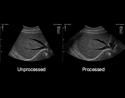
Clinical Image


Design & Ergonomics
-
01Product Specification
2 Transducer connectors (Second connector optional)
2 Built-in Batteries (Second battery optional)
Sleep mode: Max. 100 hrs.
Continuous scan: Max. 60 min.Fast System Boot-up
From sleep mode: 15 sec. / From off mode: 62 sec.15.6 inch LCD monitor
Monitor tilting up to 180°
6Kg (with 1 battery)
Dimension: 385(W) x 370(D) x 62.5~353(H) mm
4 User Define Key
Control Panel Cover (Optional)
-
02Easy control cart
Smooth and silent gas lift cylinder
Easy adjustment of height: 848 ~ 998 mm
2 transducer holders, 1 Gel holder
Weight: 22.5 kg
Safety product locks
Transducer cable management
4 swivel wheel
Safety break locks
-
03Carrying bag
3 transducer pouches included.

* The cart and carrying bag may be available for purchase separately.
Transducers
-

-
SC1-7H
X+ Crystal Signature™ Convex
ApplicationAbdomen, EM, Gynecology, Obstetrics, Pediatric, Urology
-

-
SC1-6H
High density single crystal convex transducer (1-6MHz)
ApplicationAbdomen, EM, Gynecology, Obstetrics
-

-
SC1-4H
High density single crystal convex transducer (1-4MHz)
ApplicationAbdomen, EM, Gynecology, Obstetrics
-

-
SC1-4HS
High density single crystal convex transducer (1-4MHz)
ApplicationAbdomen, EM, Gynecology, Obstetrics
-

-
C1-6CT
C-Architecture (PowerView™) Convex transducer (1-6MHz)
ApplicationAbdomen, EM, Gynecology, Obstetrics
-

-
C5-8NT
Microconvex (5-8MHz)
ApplicationAbdomen, EM, Cardiac, Pediatric
-

-
C5-8N
Microconvex (5-8MHz)
ApplicationAbdomen, Cardiac, EM
-

-
SC2-9HApplication
Abdomen, EM, Gynecology, Obstetrics, Pediatric, Urology
-

-
SC2-11H
X+ Crystal Signature™ Microconvex(2~11MHz)
ApplicationAbdomen, Pediatric, OB/GYN, Urology, EM
-

-
L8-17H
High density linear transducer(8-17MHz)
ApplicationBreast, EM, MSK, Vascular, Small Parts
-

-
L3-12X
Extreme-high density linear transducer (3-12MHz)
ApplicationBreast, EM, MSK, Vascular, Small Parts
-

-
L3-12H
High density linear transducer (3-12MHz)
ApplicationBreast, EM, MSK, Vascular, Small Parts
-

-
L3-12HWD
High density wide footprint linear transducer (3-12MHz)
ApplicationBreast, EM, MSK, Vascular, Small Parts, Appendix
-

-
L3-12T
Linear transducer (3-12MHz)
ApplicationBreast, EM, MSK, Vascular, Small Parts, Appendix
-

-
L3-8H
High density low frequency linear transducer (3-8MHz)
ApplicationBreast, EM, MSK, Vascular, Small Parts
-

-
IO3-12
Intraoral transducer (3-12MHz)
ApplicationEM , Small Parts
-

-
IO8-17T
High frequency hockey stick (8-17MHz)
ApplicationSmall Parts, MSK
-

-
SL3-19H
X+ Crystal Signature™ Linear
ApplicationAbdomen, Pediatric, Gynecology, Obstetrics, EM, MSK, Vascular, Small Parts, TCD
-

-
L10-25H
Wideband Ultra High Freq. Linear
ApplicationEM, MSK, Vascular, Small Parts
-

-
L3-15H
High density linear transducers (3-15 MHz)
ApplicationMSK, Vascular, Small Parts, EM
-

-
SL3-19X
X+ Crystal Signature™ Extreme-high density linear transducer (3-19MHz)
ApplicationAbdomen, Pediatric, Gynecology, Obstetrics, EM, MSK, Vascular, Small Parts, TCD
-

-
IO7-18
High frequency hockey stick (7-18MHz)
ApplicationSmall Parts, MSK
-

-
P1-5CT
Single crystal phased array transducer (1-5MHz)
ApplicationAbdomen, Cardiac, EM, TCD
-

-
SP3-8T
Phased array transducer (3-8MHz)
ApplicationAbdomen, Cardiac, EM, Pediatric
-

-
MP1-5X(New)
X+ Crystal Signature™ Phased Array
ApplicationAbdomen, Pediatric, Cardiac, EM, TCD
-

-
EV3-10T
Endocavity transducer (3-10MHz)
ApplicationGYN, OB, Fetal Echo, Urology, EM
-

-
EC3-10T
Endocavity transducer (3-10MHz)
ApplicationGYN, OB, Fetal Echo, Urology, EM
-

-
EV2-11H
X+ Crystal Signature™ Endocavity
ApplicationGynecology, Obstetrics, Urology, EM
-

-
EC2-11H
X+ Crystal Signature™ Endocavity
ApplicationGynecology, Obstetrics, Urology, EM
-
-
EV3-10X
Extreme high Density
ApplicationOB/GYN, Urology
-
-
EC3-10X
Extreme high Density
ApplicationOB/GYN, Urology
-

-
VE3-10H
High density volume endocavity transducer (3-10MHz)
ApplicationGYN, OB, Urology
-

-
VC1-6T
Volume Convex (1-6MHz)
ApplicationAbdomen, OB, GYN, EM
-

-
SVC1-8H
Single Crystal Volume Convex
ApplicationAbdomen, Gynecology, Obstetrics, Pediatric, Urology, EM
-

-
VE3-10H(NEW)
Volume Endocavity
ApplicationGynecology, Obstetrics, Urology, EM
-

-
CW2.0
Pencil type transducer (2.0MHz)
ApplicationCardiac
-

-
CW5.0
Pencil type transducer (5.0MHz)
ApplicationCardiac
-

-
CW8.0
Pencil type transducer (8.0MHz)
ApplicationCardiac
-

-
TEE 3-7Application
Cardiac